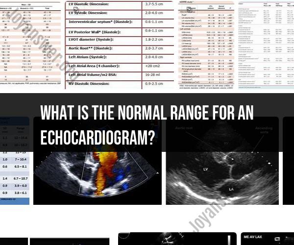
What is the normal range for an echocardiogram?
An echocardiogram, also known as an echo, is a medical test that uses ultrasound to create images of the heart's structure and function. While an echocardiogram provides detailed information about the heart, it does not have a single "normal range" like a blood test. Instead, the interpretation of an echocardiogram involves various measurements and parameters, and what's considered normal can vary depending on factors like age, sex, and individual patient characteristics.
Here are some of the key parameters and measurements that are typically assessed in an echocardiogram, along with general guidelines for what healthcare providers consider within the normal range. However, it's important to note that specific reference values can vary between different medical facilities and regions, and healthcare professionals use their judgment and expertise to interpret echocardiogram results.
Ejection Fraction (EF):
- EF measures the percentage of blood that the left ventricle pumps out with each heartbeat. A normal EF is typically between 50% and 70%. Values below 50% may indicate reduced heart function.
Left Ventricular Dimensions:
- Measurements of the left ventricle's size, including end-diastolic and end-systolic dimensions, are taken. These measurements can vary based on body size, but significant deviations may indicate heart conditions.
Valve Function:
- The echocardiogram assesses the function of heart valves, including the mitral, aortic, tricuspid, and pulmonic valves. Normal valve function means they open and close properly without significant regurgitation (backflow) or stenosis (narrowing).
Wall Motion Abnormalities:
- The test looks for any abnormalities in the movement of the heart's walls. Normal wall motion means that all parts of the heart contract and relax in a coordinated manner.
Chamber Sizes:
- Measurements of the atria and ventricles' sizes are taken. These sizes can vary depending on factors like age and body size.
Blood Flow:
- Blood flow patterns are assessed, including the direction and velocity of blood flow through the heart chambers and major blood vessels.
Pericardium:
- The echocardiogram can assess the pericardium, the sac around the heart, for any signs of fluid accumulation or inflammation.
Pressure Gradients:
- In some cases, pressure gradients across valves or chambers are measured to evaluate for conditions like valve stenosis.
Other Findings:
- Echocardiograms may also reveal other findings, such as congenital heart defects, blood clots, or tumors.
Keep in mind that echocardiogram results are interpreted by healthcare professionals, often cardiologists or cardiac sonographers, who take into account the entire clinical picture when determining whether the findings are within a normal range for the individual patient. If you have questions or concerns about your echocardiogram results, it's best to discuss them with your healthcare provider, who can provide specific information based on your medical history and condition.
Echocardiogram Normal Range: What's Considered Healthy
An echocardiogram is a non-invasive test that uses ultrasound to create images of the heart. It can be used to assess the size, shape, and function of the heart, as well as the valves and blood vessels.
The normal range for echocardiogram values varies depending on the age and gender of the patient. However, some general normal ranges include:
- Left ventricular ejection fraction (EF): 55-65%
- Left ventricular end-diastolic diameter (LVEDD): 3.5-5.6 cm
- Left ventricular end-systolic diameter (LVESD): 2.0-4.0 cm
- Right ventricular end-diastolic diameter (RVEDD): 0.7-2.3 cm
- Aortic root diameter: 2.0-4.0 cm
- Left atrial diameter: 2.0-4.0 cm
It is important to note that these are just general normal ranges. Your doctor will interpret your echocardiogram results based on your individual medical history and physical examination.
Healthy Echocardiogram Results: What to Expect
If your echocardiogram results are within the normal range, it means that your heart is likely healthy and functioning normally. However, even if your results are normal, your doctor may still recommend follow-up tests or treatment if you have other risk factors for heart disease, such as high blood pressure, high cholesterol, or diabetes.
Understanding Normal Echocardiogram Values
Here is a brief explanation of some of the key echocardiogram values:
- Left ventricular ejection fraction (EF): The EF is a measure of how well the left ventricle is pumping blood. A normal EF is 55-65%. An EF below 55% may indicate heart failure.
- Left ventricular end-diastolic diameter (LVEDD): The LVEDD is a measure of the left ventricle's diameter when it is relaxed and full of blood. A normal LVEDD is 3.5-5.6 cm. An LVEDD that is too large may indicate an enlarged heart, which can be a sign of heart failure.
- Left ventricular end-systolic diameter (LVESD): The LVESD is a measure of the left ventricle's diameter when it is contracted and empty of blood. A normal LVESD is 2.0-4.0 cm. An LVESD that is too large may indicate a weakened heart muscle.
- Right ventricular end-diastolic diameter (RVEDD): The RVEDD is a measure of the right ventricle's diameter when it is relaxed and full of blood. A normal RVEDD is 0.7-2.3 cm. An RVEDD that is too large may indicate an enlarged right ventricle, which can be a sign of pulmonary hypertension or other heart problems.
- Aortic root diameter: The aortic root diameter is a measure of the diameter of the aorta, the main artery that carries blood from the heart to the rest of the body. A normal aortic root diameter is 2.0-4.0 cm. An aortic root diameter that is too large may indicate an aortic aneurysm, which is a bulge in the aorta that can rupture and be life-threatening.
- Left atrial diameter: The left atrial diameter is a measure of the diameter of the left atrium, the chamber of the heart that receives blood from the lungs. A normal left atrial diameter is 2.0-4.0 cm. An enlarged left atrium may indicate mitral valve stenosis, a condition in which the mitral valve is narrowed and restricts blood flow from the left atrium to the left ventricle.
If you have any questions about your echocardiogram results, be sure to talk to your doctor.


