Explore the Basic Shapes
Microbes, particularly bacteria, primarily come in three distinct shapes. Each shape influences how the microbe interacts with its environment, moves, and causes disease. Click on a shape below to learn more about it.
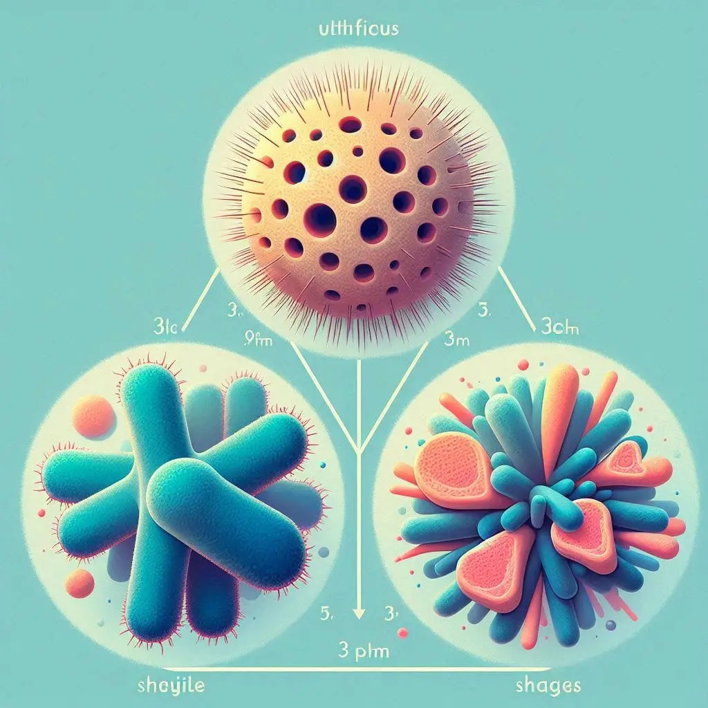
Cocci
The Spheres
Bacilli
The Rods
Spirilla
The Spirals
How We See Them: Lab Identification
Identifying a microbe's shape is a critical first step in microbiology. It's typically done through a two-part process involving microscopy and staining, which makes the transparent cells visible and provides additional information.
1. Microscopy
A sample is placed on a slide and viewed under a high-powered light microscope.
2. Staining
Dyes like the Gram stain are applied. This not only reveals the shape but also classifies bacteria based on their cell wall structure.
3. Identification
The observed shape, arrangement, and stain result provide a strong preliminary identification of the microbe.
Why Microbial Shape Matters in Medicine
Understanding the shape of a bacterium isn't just an academic exercise; it has profound implications for diagnosing and treating infectious diseases.
Rapid Diagnosis
In critical infections like meningitis, a doctor can examine spinal fluid under a microscope. Seeing spherical bacteria in pairs (diplococci) is a strong indicator of *Neisseria meningitidis*, allowing for immediate, life-saving treatment long before culture results are available.
Predicting Pathogenicity
Shape influences how bacteria cause disease. Spiral-shaped bacteria like *Helicobacter pylori* can corkscrew through the stomach's mucus lining to cause ulcers. The rod shape of *E. coli* allows for efficient formation of biofilms on surfaces.
Guiding Antibiotic Choice
The Gram stain, which reveals shape, also separates bacteria into two groups (Gram-positive and Gram-negative). This fundamental difference in cell wall structure dictates which antibiotics will be effective, making shape identification a cornerstone of treatment strategy.